 Ultrasounds are medical tests for capturing live images inside the body, very commonly used throughout the course of pregnancy. It provides the highlight of a woman’s pregnancy, such as the baby’s due date, the gender of the babyand the position of placenta. Ultrasounds enable you to check the development and health status of the baby. Many mothers recommend ultrasounds during pregnancy in order to have a positive experience throughout the various phases of pregnancy.
With the pregnancy signs being visible from the second month after you get pregnant, ultrasound scans can be carried out during any phase of pregnancy since then. Below are some of the questions which are frequently asked by many moms during pregnancy.
Is It Safe to Have an Ultrasound in Pregnancy?
Fetal ultrasound is safe. Studies have revealed that the ultrasound tests are not hazardous, and there are no harmful side effects of fetal ultrasounds to you or your baby. However, it has its own shortcomings such as providing limited information on the wellbeing of the fetus and at times, it may suggest inaccurate birth abnormalities.
Why Is an Ultrasound Done?
An ultrasound in pregnancy can make it possible to ensure on various aspects of a healthy pregnancy, such as:
- Ultrasound helps in identifying the position of your fetus during pregnancy and in diagnosing ectopic (tubal) pregnancy—the fetus develops in a fallopian tube instead of in uterus.
- Your baby’s due date can be determined through fetus ultrasound. In other words, ultrasound is used for tracking your baby’s menstrual age.
- Ultrasound helps in evaluating the size of your baby, including the development and growth of the baby. The test also confirms if you are carrying more than one baby.
- Various birth abnormalities can be detected through fetus ultrasound, and it also helps in closely monitoring the status of pregnancy, including complications like fluid loss, bleeding, and any sorts of complications with placenta.
- Ultrasound is also performed for other prenatal tests—one of the various ways to confirm on the wellbeing of your growing baby and your health status.
- Ultrasound is a helpful device used for determining the methods of childbirth—vaginal delivery or caesarean delivery, depending upon the baby’s position.
Fetal ultrasound is a boon to medical science and you shouldn’t merely go for it just to determine the baby’s gender or for keepsake pictures. Some doctors don’t recommend a fetal ultrasound, but you can still discuss with your doctor to choose the best option for your baby and yourself.
When to Have an Ultrasound in Pregnancy
|
Stages of Pregnancy |
When to Have & What to Know |
|
First Trimester |
Your initial ultrasound scan is normally initiated during 6 to 8 weeks of pregnancy in order to determine the estimated due date of the baby. This test would provide the first glimpse of the baby. Ultrasound scan is done on a full bladder as sound waves move better on liquid. |
|
Second Trimester |
The second ultrasound scan, also commonly known as anatomy scan, is generally performed during the 5th month of pregnancy. This is done to check the development and health status of the baby. The test is carried out for about 20 to 45 minutes and may prolong if you are having more than one child. Ultrasound during the second trimester can also help you in determining the gender of the baby. |
|
Third Trimester |
The final ultrasound could have been an anatomy scan which would be taken around the 20th week of pregnancy. It is normally carried out on a case by case scenarios, such as you are over 35 years of age, or your pregnancy have gone beyond your due date and your doctor wants to closely monitor your baby’s health status. |
You can also check out this video to find out more about fetus ultrasound in pregnancy:
How Do I Prepare for an Ultrasound?
It is important to know that the ultrasound scan during pregnancy is done on a full bladder as sound waves move better on liquid. Thus, you would be asked to drink at least 4 to 6 glasses of water and not to urinate before the test.
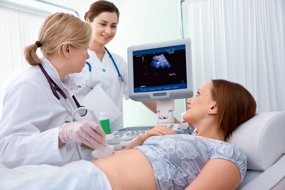
How Is an Ultrasound Performed?
Ultrasound test normally takes around 30 minutes. You would be asked to lie on your back and a water soluble gel will be put on your abdomen. A transducer, a small device is then goes backwards and forwards against the skin on your abdomen and sends high-frequency sound into the womb through your abdomen. The sound or echoes is reflected back and creates a picture on a screen. You can have the pictures printed; there may be a small charge for it. Some hospitals also allow videotaping the ultrasound scans; you may want to take the doctor’s approval for it. The lubricating gel applied on your abdomen would be wiped after the test and you can discuss the results with your doctor.
What Should I Do If the Ultrasound in Pregnancy Shows a Problem?
Ultrasound test can detect some abnormalities during pregnancy and it is very natural to get worry. Some scans may specify negligible condition like markers (minor changes) while some of the indications, such as Down’s syndrome which can be a very serious condition. You would be referred immediately to fetal medicine specialist (within 72 hours) if there is any abnormal signs during the test. Some tests such as amniocentesis or CVS are further carried out to examine the development of your baby. You will be provided the best of all the options if the test discloses a serious issue and the decision can be very difficult at times, although such complication in pregnancy is very unusual. Some problems may need you to take a special care of your baby after birth, whereas others may requisite a surgical procedure of the baby while in uterus or after birth.